
Tissue and CThree Sisters Experiment Evaluationellular Stiffening in Maladaptive Vascular Smooth Muscle Cell Phenotypic Change
The role of tissue and cellular stiffening in maladaptive vascular smooth muscle cell phenotypic change.
Aboriginal Ethnobotany
Abstract:
The three sister is a very old practice that was done by the native American in north America using corn, bean, squash. The purpose of the three sisters experiment that was conducted in the greenhouse at Lakehead University was to show whether these three crops grow better under the polyculture conditions or the monoculture conditions. The three crops were planted in a bucket that has rocks and black soil in polyculture environment and monoculture environment. Measurements were collected every week for almost 10 weeks. Measurements included soil pH, the LUX value, the plant height as well as the roots height/weight after the take down of the plants. The results have shown that the plants such as squash and corn grow better in monoculture environment rather than the polyculture environment. This contradicted the actual hypothesis that indicated that plants usually prefer growing in the polyculture environment than those that grow in the monoculture one.
Introduction
The Three Sisters crops originated in Central Mexico and were spread out around the Americas by 1000AD (Zhang et al 2014); these three sister crops consist of the cultural crop system of the Haudenosaunee (Iroquois) nation (Mt. Pleasant 2016), and are identified as the bean (Phaseolus vulgaris), squash (Cucurbita spp.) and corn/maize (Zea mays). Beans, Corn and Squash have a long history of being grown both together and apart across the American Continent; from Ontario to Florida, records show that there has been a lot of productive agriculture of these three sister crops. There are detailed reports written by Cartier supporting the cultivation of these three crops as early as 1534-1535 near present day Montreal, identifying the
Haudenosaunee agriculture as one of the earliest Indigenous crop systems to be reported by Europeans. It was initially thought by scholars that the bean, corn and squash inter-cropping system were established around 1000-1200 BP, however, paleobotanist’s John Hart’s recent research has stated that corn, beans and squash were a part of agriculture at different times. Squash was the first of the three sister crops to appear, showing up in the Northeast around the end of the third millenium, Corn appeared second around ~2270 BP New York,
Beans were the last of the sister crops to appear; they were seen to arrive in the late prehistoric period. These three crops were originally grown separate; the three sisters being grown together was observed after 700 BP (Mt. Pleasant 2016) by the Haudenosaunee nation.
If you need assistance with writing your essay, our professional essay writing service is here to help!
Corn, Beans and Squash are constantly referred to as a “symbiotic plant complex of North and Central America without an equal elsewhere”, showing the impact these three crops have when grown as a polyculture (Mt. Pleasant 2016). It is known that the Haudenosaunee first nations preferred to grow these crops as a polyculture due to it being more time and energy efficient to grow them together instead of separately, additionally there are more agricultural and dietary
benefits to growing corn, beans and squash as a polyculture. The three crops polyculture exist in harmony as per the following; Bean plants assist through the process of nitrogen fixation by maintaining a symbiotic relationship with bacteria, corn plants act as climbing poles for bean vines along with fight against weeds. Squash preserves moisture in their environment and suppress weeds, acting a lot like mulch (Hart 87). In a polyculture, these plants use soil nutrients,
light and water more efficiently and can withstand harsh climate changes better than crops grown in a monoculture (Hart 87); therefore, it is more advantageous to cultivate these three crops in a
polyculture as opposed to monocultures. Consuming corn, beans and squash together provide a more nutrient rich diet that complements each other. Corn contains a high amount of calories but low amounts of protein, lacking the essential amino acids tryptophan and lysine but having high levels of methionine. Beans compensate for corn’s lack of amino acids by containing high levels of lysine and low amounts of methionine; eating corn and beans together, increases your protein and calorie consumption. Squash is the most nutritionally rich of the three sister crops; it contains high amounts of minerals, vitamin A and calorie rich and its seeds are high in both protein and energy. (Mt. Pleasant 2016 and Hart 87). Consuming the Three Sisters together has a significantly high nutritional value, due to their vastly different nutritional contents and how they complement each other.
The objective of this scientific experiment was to familiarize ourselves and learn the benefits of polyculture versus monoculture of the three sister crops established by the Indigenous people in North America. This was carried out by comparing the following parameters; soil pH, above ground height, LUX values and root length. We hypothesized that the three sister crops; corn, beans and squash, grow more effectively in a polyculture rather than in monoculture; the polyculture will develop more structured and longer seedlings and the root systems will be longer and more developed.
Methods and materials
In this experiment, student in this lab section were divided in 5-6 groups of 4-5 students. The experiment began by each group having to develop two separate environments: monocultures of corn, bean and squash as well as a polyculture of the same selected crops. A weekly measurements were taken, and the student had the responsibility to look after the plants. The measurements include soil pH and above ground height and LUX values. As well as the root length of the crops which was done during the last week of the experiment after the take down of the plants (week 11).
The set-up of this experiment was done in the green house at Lakehead University. A 40L buckets were used for the planting of the crops. The bottom of these buckets were filled with rockets which helps provide the drainage for the root. After that, 25 L of Black Earth soil was used to fill up the buckets. The soil was split evenly for the reduction of clumps, then there was an addition fertilizer to provide nutrients for the plants.
The crops of corn, bean, squash were planted in the buckets. They were first planted in a monoculture each separately then in a polyculture together. In the monoculture environment for the squash and corn, one seedling was planted in the middle of the bucket. As well as, four seedlings were planted in square formation as shown in Figure 1. A 30 mL of all-purpose grass seedling were added to the top of soil for the stimulation of weed growth which is good for the competitions between the plants. A mesh nozzle was used for the watering of plants to prevent them from being damaged. When watering the plants, it needed to be ensured not to over water the plant as well as not to under water them. The containers were put in a place so that it receives the most sufficient amount of light.
The height of the plants was measured every week using a meter ruler. A pH probe was used to recorded the soil pH of each bucket. The LUX meter was used to record the LUX value which was done every week.
The plants were taken down out of the containers at the end of the experiment. The soil was removed from the roots as much as possible to make sure the right weight of the root is measured. The data that was obtained in the course the eleventh week was used to get the average for the making of the graphs and all the other analysis.
a) 
 b)
b)

1
1
2
3

1
4
c) d)


3
2
4
1


= corn = bean = squash
Figure 1: Illustrating the formations of the crops in each container. a) corn monoculture, b) bean monoculture c) squash monoculture and d) the Three Sisters polyculture.
Results
During the three sisters experiment, parameters such as soil pH, aboveground height, LUX values were measured weekly. additionally, before we took down the crops we measured the length of the root. The LUX values in this experiment were obtained, although it was hard to measure it most of the time as the plants started to grow.
HEIGHTS AVERAGE:
In the first week, the height of the corn in the monoculture environment was 13.3 cm, however, the height of the corn in the polyculture environment was 10.6 cm. after ten weeks, the corn in the monoculture grew significantly to 210 cm as well as the corn in the polyculture grew to 123 cm in the matter of ten weeks as shown in (figure 4). The R2 value for the line of best fit trend in the monoculture was 0.973. As for the polyculture corn, the R2 was found to be 0.921 after looking at the line of best fit (Figure 4).
Our academic experts are ready and waiting to assist with any writing project you may have. From simple essay plans, through to full dissertations, you can guarantee we have a service perfectly matched to your needs.
During the first week, the bean in the monoculture environment grew to 7.72 cm, however, in the polyculture environment the bean grew to 7.82 cm. After 10 weeks, the monoculture bean grew to become 89.3 cm in height, as well as the polyculture bean grew to 95.1 cm in height. In average the mono and poly culture bean grew 81.58 cm and 87.28 cm in height respectively (figure 2). The R2 value for the line of best fit trend in the monoculture was 0.771. As for the polyculture bean, the R2 was found to be 0.803 after looking at the line of best fit (Figure 2).
During the first week of the squash growth, the monoculture squash had barley in growth, this was the case for the polyculture squash as well. During the third week, there was a growth of 17.6 cm in the monoculture squash and a growth of 18.3 cm in the polyculture squash. In the last week of the experiment, the mono squash grew to 91 cm, however the poly squash grew to 68 cm in height. In average both mono and poly squash had a growth of 58.4 and 54.4 cm respectively (figure 3). The R2 value for the line of best fit trend in the monoculture was 0.888. As for the polyculture bean, the R2 was found to be 0.771 after looking at the line of best fit (Figure 3). By and large, the plants in the polyculture environment had a long growth in height when compared to the plants in the monoculture environment. With to the exception to the bean crops where the monoculture crops had a lager growth in height in comparison to the polyculture.
Soil pH
Figure 4 shows the average of the soil pH of the plant cultures in the period of ten weeks. The values that was obtained varied from 4 to 7. During the first weeks, there was no major changes and it seems very stable, however there were changes in the pH during third, forth, fifth and sixth week. The pH in the last weeks 7-11 was slightly stable and there were no major changes in it (figure 5).
ROOT LENGTH
Before the take down of the crops, a measurement of the root length needed to be done. In the monoculture environment, the average length of the bean was around 110.6 cm, whereas the polyculture bean has an average length of 138.6 cm (Figure 6). The length difference in the mono and poly bean was around 28 cm.
In the corn monoculture environment, the corn had a growth length average of 120 cm, however, the corn in the polyculture environment had a growth length average of 110 cm. The corn in the monoculture environment grew around 10 cm longer that the polyculture one (Figure 6).
In the Squash monoculture environment, the squash had a growth length average of 91 cm, while, the squash in the polyculture environment had a growth length average of 68 cm. the difference in both culture was 23 cm for the monoculture squash. (Figure 6). Figure 5 shows a trend of the crops in the monoculture environment growing longer than the crops that grew in the polyculture environment.

Figure 2: Comparison of average above ground heights of the bean monoculture and the crops in the polyculture in the none-week period.

Figure 3: Comparison of the average above ground heights of the squash monoculture and the crops in the polyculture in the nine- week period.

Figure 4: Comparison of the average above ground heights of the crops in the corn monoculture polyculture in the nine-week period.

Figure 5: Comparison of the weekly average soil pH of bean, corn, squash and the polyculture.
The pH of the soil of the four cultures was measure once every week by each group.

Figure 6: Comparison of the average root lengths of the crops in the monocultures and the
polyculture at the end of nine-week period.
Discussion:
There have been many studies that were conducted regarding the three sister topic. Most the results showed that the polyculture is a better environment for the growth of plants because it has a greater biomass as well as a higher yield than those that grow in the monoculture. (Zhang et al 2014). A statement that was made by Zhang et al. stated that the biomass, the nutrient uptake as well as the yield in various soils were the reasons that the polycultures are more effective and valuable. (Zhang et al 2016). One of the reasons that The Three Sisters crops, corn, bean and squash polyculture was successful is that these three crops are very complementing to each other. Corns provide a climbing pole for the bean and it also works as a competitive factor against all weeds. Squash plays a good role for the preservation of the soil as well as it helps supressing the weed. Squash also is used for the nitrogen fixation in the bean plant in the atmosphere. (Hart 87). However, bean is a very good supply of nitrogen, it supplies about 20-60% of nitrogen to the soil which is done through symbiotic nitrogen fixation. Then corn and bean uses the soil for their nitrogen’s intake. (Postma 2012). The Three Sisters are very complementing amongst each other in the polyculture environment by their spatial segregation and soil resource partitioning. This might be due to the difference in the root system. In the bean, the basal roots and tap is what control the root system, whereas the corn has a nodal root system. Furthermore, the tap root and larger lateral is very dominant in the squash. Therefore, the different roots in these three crops utilize different areas and layers in the soil which explains why they are very complementing to each other. As a result of that, a segregation in a blew-ground spatial niche will occur. (Zhang et al 2014). Polyculture in the three sisters experiment are more successful than growing the crops in the monoculture environment based on many reasons that some of it was discussed in this report.
The hypothesis in this experiment was that, although the crops in the polyculture tend to grow more successfully with a more developed root system comparing to those that grow in the monoculture environment, some of the data that was obtained and measured throughout the eleventh weeks have shown otherwise. The bean has met the expectation where its crops grew way more effectively in the polycultures as a pose to the monocultures. Whereas the corn and squash contradicted the hypothesis because they grew better in the monoculture. One of the reasons might be because of the lack of fertilizer between both cultures. Equivalent amounts of fertilizer were given to each container, causing the polyculture plants to share between each other as opposed to the monoculture plants, who did not. The average seedling height in the final week was larger in the squash replicates who were in the polyculture, as the seedlings planted were longer than those of an average monoculture squash seedling, allowing them to start at a longer height and thus, have a larger final height at the finish of this experiment. Additionally, the corn and bean seedlings planted in polyculture were longer on average than their monoculture seedling counterparts. However, it was observed that monocultures starting at shorter lengths grew longer than their polyculture counterparts, meaning, the polyculture plants were not very successful. Errors occurred in this experiment that resulted in skewed results which will be discussed further on in this report. The pH measurements that resulted from this experiment did not show any specific trend; values fluctuated from 4-7.5 (Table 2). pH fluctuations are a common occurrence in soil, when plants are growing, roots take up many nutrients from soil (Hinsinger et al 2003); more nutrients in soil causes it to be more acidic. Roots ingest nutrients according to nutritional requirements causing soil to be more basic; this explains the pH fluctuations that occurred over the 11-week period. Our hypothesis stated that root lengths of polyculture crops would be longer than their monoculture counterpart; however, the experimental results showed otherwise. Our results are as follows; monoculture plants grew longer roots than their polyculture counterparts as seen in Figure 5. This is due to the polyculture crops replicating and growing in clusters, in a small space. All the replicates were grown in 40 L containers therefore, the polyculture crops did not have enough space to spread out roots and grow properly, additionally, squash and corn did not suppress the weeds as expected. The monocultures had more space to grow their root system and develop more and stronger. If they had a bigger experimental area, the polyculture crop roots would compartmentalize the soil according to their root system, thus, they would no longer have to compete with each other below-ground. These three crops are able to compartmentalize soil, creating niche segregation as corn has a nodal root, bean has a tap and basal root and squash has a tap and a lateral root system as stated earlier in this discussion (Zhang et al 2014).
There have been many errors that have happen during the performance of this experiment which might have caused in some of the results meeting the actual results. One of the factors might have been the way our group looked after the plants. Some of the weeks the plants were not watered enough and other times the plants were over watered. This could have resulted in the plants being overly dried and not being able in the conditions that they acquire. During the experiment, the plants were supposed to be sprayed for the prevention of the aphid. This aphid can cause a decrease in the plants’ growth by putting stress on them. The light that the plants was supposed to use is another factor why the results were not very ideal since there was not a lot of sun coming through the greenhouse for the plants to get exposed to. Also, as the data was collected there was probably mistakes that happened since every time a different student would go alone and measure everything as a posed to the whole group being in attendance.
There many things that can be done for the improvement of this experiment in the future. A lager buckets could be used to provide a greater the space for the growth of the plants in the polyculture since a smaller area would cause a bigger competition amongst the crops. A better watering technique could be used for the prevention of the over watering of the plants which can damage it. The LUX can be measured differently and each group can have the ability to come in different time during the day to obtain more ideal measurements. Additionally, looking at the food yield form the plants could be helpful instead of only comparing the height of the plants only. These are some of the suggestions that might help making the more successful in the future.
Conclusion:
To conclude, the three sisters crops is an excellent technique that was used by indigenous people long time ago. The results in most of the experiment that were done in the past showed that the polycultures are a better environment for the three crops to grow, however the results that was obtained during this experiment showed that the monocultures were a better condition. This might be due to the errors that might have been done throughout all week. Avoiding some of the mistakes that was mentioned in the paper will results in a better project.
References
- Hart, John., Evolving the Three Sisters: The Changing Histories of Maize, Bean, and Squash in New York and the Greater Northeast. pp. 87.
- Hinsinger, Philipper., Plassard, Claude., Tang, Caixian., Jaillard, Benoit. 2003. Origins of root-mediated pH changes in the rhizosphere and their responses to environmental constraints: a review. Plant and soil. 248(1-2), pp. 43-59.
- Mt. Pleasant, J., 2016. Food yields and Nutrient Analyses of the Three Sisters: A Haudenosaunee Cropping System. Ethnobiology Letters. 7(8), pp. 87-98.
- Postma, J. A., Lynch, J.P., 2012. Complementarity in root architecture for nutrient uptake in ancient maize/bean and maize/bean/corn polycultures. Annals of botany. 110, pp. 521-534.
- Zhang, C., Postma, J.A., York, L.M. and Lynch, J.P., 2014. Root foraging elicits niche complementarity-dependent yield advantage in the ancient ‘three sisters’(maize/bean/squash) polyculture. Annals of botany. 114(8), pp.1719-1733
- Cardiovascular Disease
Cardiovascular disease (CVD) refers to a heterogeneous group of diseases afflicting the heart and vasculature with similar underlying pathophysiological manifestations, such as coronary heart diseases, cerebrovascular disease, hypertension, peripheral arterial disease, congenital heart diseases and heart failure. The prevalence of CVD had reached global epidemic proportions, with an estimated 422.7 million cases worldwide [Figure 1]1. CVD is the universal leading cause of mortality, resulting in 17.9 million deaths in 2015, accounting for 44% of all non-communicable disease deaths2. In the UK, around 7 million people are living with CVD and 25% of all deaths (>150,000 deaths/annum) are attributable to CVD3.
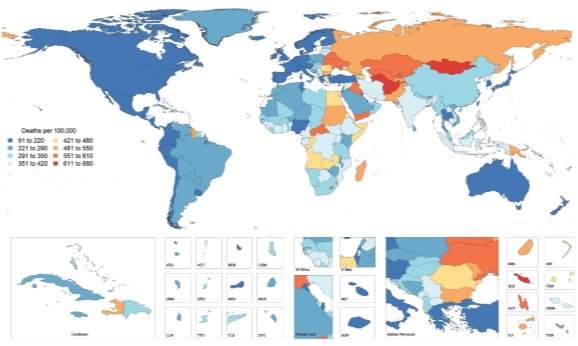
Figure 1: Global Map of Age-Standardised Death Rate of CVD in 2015. Choropleth map illustrating the estimated age-standardized mortality rate of total CVD in 2015 for each country1.
The worldwide burden of CVD on public health is escalating due to global demographic and socio-economic trends, particularly population ageing, economic growth in Asia and the obesity epidemic observed in developed countries. In addition, more than 75% of CVD related deaths occur in low- and middle-income countries, as a consequence of restricted access to primary healthcare and affordable treatments. Thus, an enhanced understanding of cardiovascular physiology and CVD mechanisms, as well as the development of novel, more targeted and low-cost prevention, early diagnostic and therapeutic strategies are required to reduce the incidence of CVD mortality4.
If you need assistance with writing your essay, our professional essay writing service is here to help!
CVD pathogenesis and related complications are attributed to numerous risk factors including metabolic factors like hypertension, hyperglycaemia, dyslipidaemia and obesity, coupled with behavioural factors such as imbalanced diet, physical inactivity, excessive alcohol consumption and tobacco use. Ageing is an integral, independent risk factor for CVD5, however many questions are unanswered concerning ageing and CVD. Here, the focus will be on elucidating possible links between ageing, CVD and vascular calcification.
- Ageing
- Arterial Stiffening
Arterial compliance is dependent upon intrinsic material stiffness and the arterial architecture. Arterial stiffness can be defined as a diminishing distensibility that characterises the relative changes occurring in the lumen cross-sectional area for a given variation in blood pressure18.
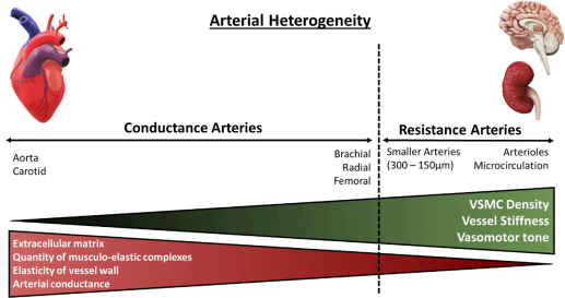
The distensibility or stiffness of the arteries contributes to wave propagation and reflection in the arterial vasculature, where the arterial pulse disseminates with a certain speed known as the pulse wave velocity (PWV), continuously varying in amplification and shape. Arterial stiffness has aetiological implications in various cardiovascular diseases and has been identified as a key independent risk factor for all-cause and CVD mortality19,20. Aortic PWV is a reference parameter largely accepted as the simplest, non-invasive, accurate and reproducible measurement of central arterial stiffness for large elastic arteries. Clinically, the carotid-femoral PWV is considered as the ‘gold-standard’ measurement to determine arterial stiffness, measured along the aorto-iliac pathway and has been previously utilised to determine the predictive value of aortic stiffness and cardiovascular incidence21. Arterial stiffness indexes, for example, Young’s elastic modulus calculated from stress-strain curves, are utilised for smaller-sized muscular arteries. Both central and peripheral stiffness is influenced by the intricate interactions of the intramural cells and ECM, which are responsible for the mechanical function and structural integrity of the vasculature, differing with vessel size22. The aetiology of arterial stiffening is attributed to various molecular and cellular determinants including key ECM components (primarily elastin and collagen fibres), cell-ECM interactions and VSMC tone regulatory proteins. In addition, mechanical factors of arterial wall remodelling (shear stress and pulsatile, circumferential stress) and VSMC plasticity are key determinants of arterial stiffness18.
Figure 2: Architectural and functional divergence of the arterial vasculature. The diversity of the arterial vasculature ensures that conductance and resistance arteries can exert their respective functions. Larger elastic arteries, known as conductance arteries, convert pulsatile flow and pressure into continuous pressure, enabling oxygen delivery with nominal energy loss in arteriolar vascular walls. Resistance arteries are smaller in diameter, include arterioles and the microcirculation, and are responsible for the distribution of blood flow to target organs. Red triangle: Demonstrates arterial compliance of various arteries, amount of ECM present within vessels’ wall and number of musculo-elastic complexes. Green triangle: Demonstrates density of VSMCs within arterial wall, vessel stiffness and vasomotor tone, the function of which augments with diminishing vessel size. Adapted from Lacolley, P. et al (2017)14.
- Smooth muscle cells
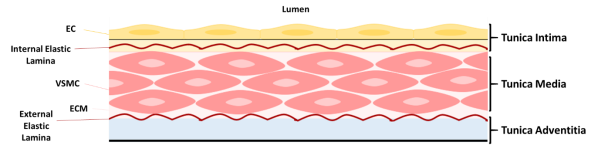
Smooth muscle is a non-striated, involuntary muscle that is distributed mainly in the hollow and tubular organs, for example, bladder, trachea, digestive tract and the vasculature. Smooth muscle consists of mononucleated, elongated and fusiform cells, which produce copious amounts of extracellular matrix (ECM)6. The principal function of smooth muscle cells (SMCs) is to contract and produce mechanical output required for specific organ function. Vascular smooth muscle cells (VSMCs) are stromal, contractile cells and the predominant cell type found in the tunica media of blood vessels [Fig.2]7. In addition, VSMCs maintain and remodel the ECM in the vasculature8.
Figure 3: Structure of blood vessel wall. The tunica intima consists of endothelial cells (ECs) resting on the internal elastic lamina. The tunica media comprises of elastin and collagen fibres and loosely dispersed, circumferentially aligned vascular smooth muscle cells (VSMCs) in extracellular matrix (ECM), embedded between the internal and external elastic laminae. VSMCs are attached to the elastic laminae by dense plaques, converging to focal adhesion complexes. Collagen fibres occur laterally to the elastic laminae. Oxytalan fibres, composed of fibrillin, connect VSMCs to the elastic laminae. The tunica adventitia is composed of collagen fibres and fibroblasts. Adapted from Lacolley, P. et al (2017)and Ponticos, M and Smith, B (2014).
VSMCs demonstrate a diverse mosaic developmental pattern, deriving from various embryogenic tissues such as the proepicardium, mesothelium, neural crest and somites9. Flk1-positive embryonic stem cells serve as vascular progenitors, for endothelial cells and perivascular mural cells, including VSMCs and pericytes10. The primary function of VSMCs is to provide structural stability, functionality and to resist mechanical stress in the vasculature effectuated by the high-pressure load of circulating blood. Through coordinated contraction and relaxation cycles in opposition to the heart, VSMCs adjust vessel diameter, regulate haemodynamics and prevent physical permeability in large vessels, ensuring mechanical compliance and sustaining normal blood pressure11,12.
4.1. Smooth muscle contraction and relaxation
SMC contraction/relaxation signalling and mechanics are coordinated by the 20kDa myosin light chain protein (MLC20). Active MLC20 stimulates myosin Mg2+ ATPase function, enabling its binding and sliding along the actin filament, concluding in cell and muscle contraction. There are two main contraction signalling mechanisms in SMCs. The Ca2+-calmodulin system, which requires Ca2+-dependent activation of MLC20 and is mediated by myosin light chain kinase (MLCK). An influx of extracellular Ca2+ through plasmalemmal Ca2+ channels and the release of sarcoplasmic reticulum Ca2+ precipitates in an incline of free cytoplasmic Ca2+, resulting in the activation of the Ca2+-calmodulin system. Calmodulin and intracellular Ca2+ form complexes, activating MLCK and initiating SMC contraction. Vasoactive ligands include vasopressin, angiotensin II, endothelin-1, norepinephrine and prostaglandins.
Our academic experts are ready and waiting to assist with any writing project you may have. From simple essay plans, through to full dissertations, you can guarantee we have a service perfectly matched to your needs.
Ca2+-sensitisation is the second contraction signalling mechanism. It is Ca2+ independent, facilitated by the RhoA/Rho-associated protein kinase (ROCK) pathway and its primary function is the maintenance of force generation in the absence of adequate intracellular Ca2+ concentrations. RhoA recruits and activates ROCK, which in turn, phosphorylates MLC phosphatase and MLC20, stimulating SMC contraction. Relaxation is accomplished through the elimination of contractile stimuli, for example, the decline of free cytoplasmic Ca2+ and/or the direct action of vasodilator agonists, including natriuretic peptides, nitric oxide, insulin and adenosine13–16.
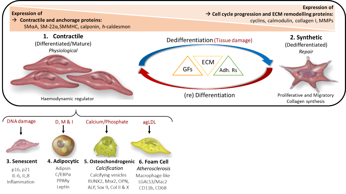
4.2. VSMC Plasticity
Under normal physiological conditions, mature VSMCs are phenotypically contractile and differentiated, with a slow proliferation rate. They respond to stimulatory neurotransmitters (e.g. acetylcholine) and express various contractile and anchorage markers including smooth muscle α-actin (SMαA), SM-22α, SM myosin heavy chains SM-1, SM-2 (SMMHC-1,-2) calponin, h-caldesmon and smoothelin17. However, VSMCs are not terminally differentiated and display functional phenotypic plasticity, which is exhibited in response to vascular injury or local environmental signals (e.g. paracrine signals, cell-cell and/or cell-matrix interactions). As a result of tissue damage, VSMCs modulate their phenotype and acquire functional properties such as migratory, synthetic and proliferative, which in turn, facilitate tissue repair.
Repair and renewal : Mature VSMCs contribute to smooth muscle and vessel repair due to their enhanced cellular plasticity, facilitating phenotypic switching from differentiated (contractile-quiescent) to dedifferentiated (synthetic and proliferative-migratory).
- Vascular Calcification
VSMCs are the fundamental cell type implicated in vascular calcification.
Points to write about:
- Talk about morphological changes observed in aorta of young and aged rats (Abu-Dief (2016))
References ADD URL and Date for WHO and BHF references
- Roth, G. A. et al. Global, Regional, and National Burden of Cardiovascular Diseases for 10 Causes, 1990 to 2015. J. Am. Coll. Cardiol. 70, 1–25 (2017).
- WHO. Cardiovascular Disease Statistics. (2017).
- BHF. British Heart Foundation UK Factsheet (BHF estimate based on latest Quality & Outcomes Framework prevalence data). (2018).
- World Health Organization. World health statistics. 2016, Monitoring health for the SDGs, Sustainable Development Goals.
- North, B. J. & Sinclair, D. A. The intersection between aging and cardiovascular disease. Circ. Res. 110, 1097–108 (2012).
- Kao, C., & Carsten, M. (Eds. . Cellular Aspects of Smooth Muscle Function. (Cambridge University Press, 1997). doi:10.1017/CBO9780511759383
- Metz, R. P., Patterson, J. L. & Wilson, E. Vascular smooth muscle cells: Isolation, culture, and characterization. Methods Mol. Biol. (2012). doi:10.1007/978-1-61779-523-7_16
- Ponticos, M. & Smith, B. D. Extracellular matrix synthesis in vascular disease: hypertension, and atherosclerosis. J. Biomed. Res. 28, 25–39 (2014).
- Majesky, M. W., Dong, X. R., Regan, J. N. & Hoglund, V. J. Vascular smooth muscle progenitor cells: building and repairing blood vessels. Circ. Res. 108, 365–77 (2011).
- Yamashita, J. et al. Flk1-positive cells derived from embryonic stem cells serve as vascular progenitors. Nature 408, 92–96 (2000).
- Frismantiene, A., Philippova, M., Erne, P. & Resink, T. J. Smooth muscle cell-driven vascular diseases and molecular mechanisms of VSMC plasticity. Cell. Signal. 52, 48–64 (2018).
- Dart, A. M. & Kingwell, B. A. Pulse Pressure-A Review of Mechanisms and Clinical Relevance. J. Am. Coll. Cardiol. 37, 975–984 (2001).
- Touyz, R. M. et al. Vascular smooth muscle contraction in hypertension. Cardiovasc. Res. 114, 529–539 (2018).
- Eto, M. & Kitazawa, T. Diversity and plasticity in signaling pathways that regulate smooth muscle responsiveness: Paradigms and paradoxes for the myosin phosphatase, the master regulator of smooth muscle contraction. doi:10.1540/jsmr.53.1
- Brozovich, F. V et al. Mechanisms of Vascular Smooth Muscle Contraction and the Basis for Pharmacologic Treatment of Smooth Muscle Disorders. Pharmacol. Rev. 68, 476–532 (2016).
- Berridge, M. J. Smooth muscle cell calcium activation mechanisms. Journal of Physiology (2008). doi:10.1113/jphysiol.2008.160440
- Durham, A. L., Speer, M. Y., Scatena, M., Giachelli, C. M. & Shanahan, C. M. Role of smooth muscle cells in vascular calcification: Implications in atherosclerosis and arterial stiffness. Cardiovascular Research (2018). doi:10.1093/cvr/cvy010
- Lacolley, P., Regnault, V., Segers, P. & Laurent, S. Vascular Smooth Muscle Cells and Arterial Stiffening: Relevance in Development, Aging, and Disease. Physiol. Rev. 97, 1555–1617 (2017).
- 19. Ben-Shlomo, Y. et al. Cardiometabolic Risk Aortic Pulse Wave Velocity Improves Cardiovascular Event Prediction An Individual Participant Meta-Analysis of Prospective Observational Data From 17,635 Subjects. J. Am. Coll. Cardiol. 63, 636–646 (2014).
- 20. Laurent, S. et al. Aortic Stiffness Is an Independent Predictor of All-Cause and Cardiovascular Mortality in Hypertensive Patients. Hypertension 37, 1236–1241 (2001).
- 21. Laurent, S. et al. Expert consensus document on arterial stiffness: methodological issues and clinical applications on behalf of the European Network for Non-invasive Investigation of Large Arteries. Eur. Heart J. 27, 2588–2605 (2006).
- 22. Humphrey, J. D., Dufresne, E. R. & Schwartz, M. A. Mechanotransduction and extracellular matrix homeostasis. Nat. Rev. Mol. Cell Biol. 15, 802–812 (2014).